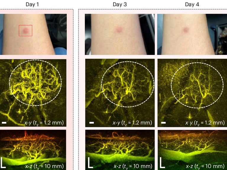
A breakthrough in miniaturising a type of scanner that uses laser light, rather than harmful X-rays, to see beneath our skin in unprecedented detail may help revolutionise medical imaging, according to a team of researchers in the UK.
The device, developed by scientists at University College London (UCL), is particularly effective at imaging blood vessels, making it a fundamentally new tool for diagnosing and managing diseases like arthritis, diabetes and some cancers.
It uses a technique called Photoaccoustic Tomography (PAT) that uses laser light, and the ultrasound waves it triggers in certain tissues, to piece together a three-dimensional image of our biology in real-time.
The technique was pioneered more than 20 years ago, but previous versions required several seconds or minutes to record an image.
The team at UCL have reduced that time to a second or less.

They hope their breakthrough will lead to a hand-held scanner for routine use in clinics that avoids the use of harmful X-rays or multi-million pound imaging tools like MRI.
"These technical advances make the system suitable for clinical use for the first time, allowing us to look at aspects of human biology and disease that we haven't been able to before," said Professor Paul Beard, a medical physicist at UCL who contributed to the research.
The speed at which the scanner can take images allows the device to see processes like blood-flow in real time.
"This speed avoids motion-induced blurring, providing highly-detailed images of a quality that no other scanner can provide," said Prof Beard.
"It also means that rather than taking five minutes or longer, images can be acquired in real time, making it possible to visualise dynamic physiological events."
A trial of the scanner involving patients with early-stage diabetes revealed new insights about low blood flow to their feet - one of the most painful and hard to treat aspects of the condition.
"Until now we haven't been able to see exactly what is happening to cause this damage or characterise how it develops," said Andrew Plumb, one of the study authors and associate professor of medical imaging at UCL.
"In one of our patients, we could see smooth, uniform vessels in the left foot and deformed, squiggly vessels in the same region of the right foot, indicative of problems that may lead to tissue damage in future."
Read more from science and technology:
Inside the UK's first fossil fuel free flying school
Elon Musk lashes out after 'summit snub'
'Mini-moon' about to enter Earth's orbit
Keep up with all the latest news from the UK and around the world by following Sky News
Tap hereThe hope is a PAT-scanner could also improve the diagnosis and treatment cancer.
Cancer tumours often have a high density of small blood vessels that don't show up well using other imaging techniques.
"Photoacoustic imaging could be used to help cancer surgeons better distinguish tumour tissue from normal tissue by visualising the blood vessels in the tumour, helping to ensure all of the tumour is removed during surgery and minimising the risk of recurrence," said Dr Nam Huynh from UCL Medical Physics and Biomedical Engineering who helped develop the scanner.
The UCL team say more work is needed with a larger group of patients to demonstrate the potential of the technology before it is ready to be developed into a device for routine use in clinics.
Disclaimer: The copyright of this article belongs to the original author. Reposting this article is solely for the purpose of information dissemination and does not constitute any investment advice. If there is any infringement, please contact us immediately. We will make corrections or deletions as necessary. Thank you.


